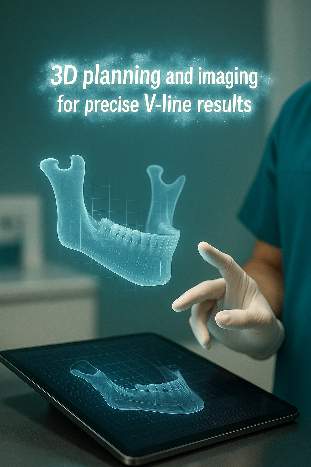
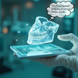
3D planning and imaging for precise V-line results is the cornerstone of modern mandibular contouring. By integrating high-resolution imaging, virtual surgical planning (VSP), and patient-specific guides, surgeons minimize intraoperative guesswork and maximize accuracy. This article delves into the technical infrastructure, imaging modalities, software platforms, guide fabrication methods, accuracy metrics, and quality-control steps that define a state-of-the-art 3D planning and imaging for precise V-line results protocol.
-
آنچه در این مقاله میخوانید
Imaging Modalities: Foundation of Accurate Anatomy
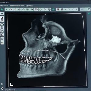
The first step in 3D planning and imaging for precise V-line results is acquiring volumetric anatomical data. Two principal modalities are used:
• Cone-Beam Computed Tomography (CBCT): Offers sub-millimeter resolution of craniofacial bone with low radiation dose. Ideal for capturing mandibular angles and chin morphology.
• Multi-Slice Computed Tomography (MSCT): Provides superior soft-tissue contrast and finer bony detail when needed for complex osteotomies.
In either case, data are exported in DICOM format for segmentation. Optimized imaging protocols—including 0.2–0.4 mm slice thickness, artefact reduction filters, and patient stabilization—ensure the raw data support 3D planning and imaging for precise V-line results.
2. Segmentation and Virtual Anatomical Modeling
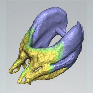
Once DICOM data are imported, 3D planning and imaging for precise V-line results relies on precise segmentation:
1. Thresholding: Automatic bone segmentation uses Hounsfield unit thresholds to delineate cortical and cancellous bone.
2. Region-growing and manual editing: Ensures the mandibular body, ramus, and symphysis are accurately isolated.
3. Mesh smoothing and error correction: Eliminates scanning artefacts and non-manifold geometry, producing a watertight 3D model.
Accurate segmentation underpins all subsequent steps in 3D planning and imaging for precise V-line results, as errors here propagate through the workflow.
3. Virtual Surgical Planning (VSP)
Virtual surgical planning is the heart of 3D planning and imaging for precise V-line results. Key components include:
• Osteotomy design: Interactive tools allow the surgeon to define bone cuts for mandibular angle reduction and genioplasty.
• Simulated bone repositioning: Real-time rendering shows how resected and repositioned segments impact lower-face contour.
• Soft-tissue prediction models: Biomechanical algorithms estimate postoperative soft-tissue drape, refining the surgical plan.
• Cephalometric analysis integration: Traditional angular and linear measurements (e.g., gonial angle, pogonion projection) guide digital manipulations.
The iterative VSP process is repeated until the desired 3D planning and imaging for precise V-line results is achieved, combining surgical feasibility with aesthetic goals.
4. CAD/CAM Surgical Guide Fabrication
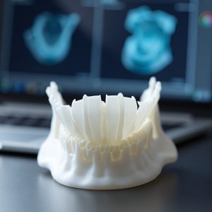
Translating the VSP into the operating room requires patient-specific instruments:
• Guide design: Using the final virtual plan, CAD software generates cutting and repositioning guides that fit the patient’s mandibular surface.
• Material selection: Biocompatible, sterilizable polymers (e.g., nylon via selective laser sintering) ensure guides maintain dimensional stability.
• 3D printing accuracy: High-resolution printers (layer thickness ≤ 50 µm) reproduce guide geometries to within ±0.1 mm, critical for 3D planning and imaging for precise V-line results.
• Post-processing: Supports removal, sterilization checks, and fit testing on 3D-printed mandibular models.
These guides enable surgeons to execute osteotomies exactly as designed in the virtual environment.
5. Intraoperative Navigation and Verification
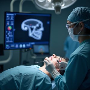
Some advanced centers combine guide use with optical or electromagnetic navigation:
• Registration: Fiducial markers or surface-mapping techniques align the patient’s anatomy with the virtual model.
• Real-time tracking: Surgical instruments are monitored relative to the registered anatomy, allowing verification of guide placement and cut angles.
• Accuracy assessment: Intraoperative CBCT or fluoroscopic checks confirm that bone resections adhere to the plan.
Integration of navigation into 3D planning and imaging for precise V-line results further reduces variability and enhances reproducibility.
6. Quality-Control Metrics
Outcome fidelity is monitored by comparing preoperative plans with postoperative reality:
• Superimposition analysis: Postoperative CBCT is overlaid onto the original virtual plan. Deviations at key landmarks (gonion, menton) are quantified; acceptable error is typically < 1 mm.
• Angular consistency: Changes in gonial angle and mandibular plane angle are measured against planned values.
• Soft-tissue correspondence: Photogrammetric or 3D stereophotogrammetry systems assess whether soft-tissue changes align with predicted models.
Regular audit of these metrics completes the 3D planning and imaging for precise V-line results feedback loop, informing continuous improvement.
7. Software Ecosystem
A robust 3D planning and imaging for precise V-line results workflow demands interoperable software:
• Medical-grade DICOM viewers (e.g., Mimics, Dolphin 3D) for segmentation and basic analysis.
• Dedicated VSP platforms (e.g., IPS CaseDesigner, ProPlan CMF) offering osteotomy and simulation tools.
• CAD software (e.g., 3-matic, Geomagic Freeform) for guide design.
• Navigation systems (e.g., Brainlab, Stryker) enabling intraoperative tracking.
Choosing validated, FDA-approved solutions ensures data integrity and regulatory compliance.
8. Team Collaboration and Workflow Integration
Successful 3D planning and imaging for precise V-line results depends on multidisciplinary collaboration:
• Surgeons define anatomical targets and aesthetic endpoints.
• Biomedical engineers handle segmentation, guide design, and printing.
• Anesthesiologists plan intraoperative imaging and navigation support.
• Nursing and technical staff manage sterilization, device checks, and intraoperative logistics.
Clear communication protocols and version control of digital files are essential to avoid errors in 3D planning and imaging for precise V-line results.
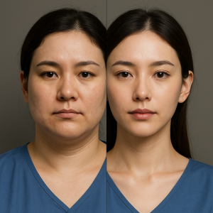
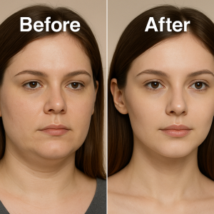
9. Case Example

A 28-year-old female presented with a broad mandibular angle and mild chin retrusion. Following 3D planning and imaging for precise V-line results:
1. CBCT segmentation revealed a gonial angle of 130°.
2. VSP targeted reduction to 120° and 3 mm chin advancement.
3. Guides were fabricated and printed to ±0.1 mm tolerance.
4. Intraoperative navigation confirmed guide placement.
5. Postoperative CBCT showed 121° and 2.8 mm advancement (error < 0.5 mm).
This precision translated into a smooth, feminine V-line with high patient satisfaction.
10. Challenges and Limitations
While 3D planning and imaging for precise V-line results offers unprecedented accuracy, limitations exist:
• Segmentation errors from artefacts or patient movement can misrepresent anatomy.
• Soft-tissue variability means predictions are approximations, not certainties.
• Cost and resource demands for equipment and trained personnel limit availability.
• Learning curve for surgeons and engineers in mastering digital tools impacts workflow efficiency.
Understanding these factors is key to realistic expectations and risk management.
11. Future Directions
Emerging technologies promise to refine 3D planning and imaging for precise V-line results further:
• AI-driven segmentation reduces manual editing time and improves consistency.
• Augmented reality (AR) overlays project the virtual plan directly onto the surgical field.
• Bioprinting of patient-specific bone scaffolds for reconstruction and augmentation.
• Cloud-based collaboration platforms enable remote planning across centers, democratizing access.
These innovations will continue to push the envelope of precision in V-line contouring.
3D planning and imaging for precise V-line results represents a paradigm shift in mandibular contouring. By integrating advanced imaging, virtual simulation, CAD/CAM guides, and intraoperative navigation, surgeons in Tehran achieve reproducible, high-fidelity outcomes. As digital workflows evolve, continuous audit of accuracy metrics and multidisciplinary collaboration ensure that each patient receives a meticulously planned and executed V-line transformation.
For international patients seeking world-class digital precision in V-line surgery, Dr. Mani Arash Rad’s clinic in Tehran offers the full spectrum of 3D planning and imaging for precise V-line results, combining cutting-edge technology with surgical expertise.
Frequently Asked Questions (FAQs)
What is the role of 3D planning and imaging for precise V-line results?
3D planning and imaging for precise V-line results provides a virtual roadmap—using CBCT or MSCT data—to design osteotomies and simulate soft-tissue changes before ever touching bone, ensuring accuracy and predictability.
Which imaging modality is best for virtual surgical planning?
Cone-beam CT (CBCT) is preferred for mandible contouring due to its sub-millimeter resolution and low radiation. Multi-slice CT (MSCT) may be used when additional soft-tissue detail is needed.
How do patient-specific surgical guides improve outcomes?
Guides fabricated via CAD/CAM from the virtual plan fit the jaw precisely, translating digital cuts into the OR with <0.1 mm error—central to 3D planning and imaging for precise V-line results.
Can I visualize my expected jawline before surgery?
Yes. Virtual surgical planning software lets you preview mandibular angle reductions and chin reshaping, offering a clear before-and-after comparison as part of 3D planning and imaging for precise V-line results.
Is intraoperative navigation necessary?
Not always, but when combined with surgical guides, optical or electromagnetic navigation adds an extra verification layer, further reducing variability in 3D planning and imaging for precise V-line results.
Ready to experience the highest level of precision in your V-line transformation? Contact Dr. Mani Arash Rad’s team for a dedicated 3D planning and imaging for precise V-line results consultation and secure your advanced digital workflow appointment today.
📍 Clinic: Saadat Abad, Sina Medical Center, Tehran
📱 WhatsApp: +98 919 789 0709